Application
Neuro & Point-of-Care
Head imaging at the bedside, in the ward, and in the operating room — reducing transport risk for critical patients.
Why it works
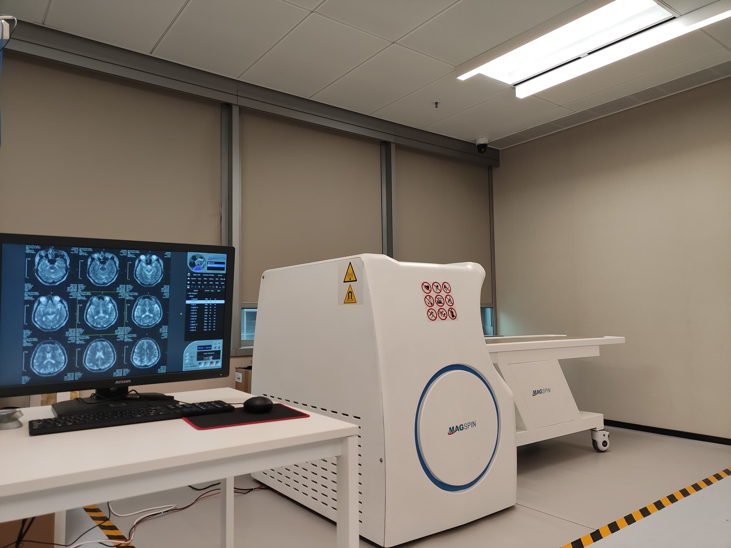
The setup.
Head imaging is where access is hardest to arrange and the cost of waiting is highest. A conventional scanner is fixed, usually on a lower floor, and reaching it means moving a patient who may be unstable, monitored, or unable to tolerate a closed bore at all.
A transportable system with an open geometry moves the imaging rather than the patient. The study happens where the care is happening — ward, emergency department, or operating room — and the tunnel that makes a share of patients need sedation disappears along with the move.
- A small modular enclosure rather than a full shielded room.
- Single-phase power and, in most sites, no floor reinforcement.
- Permanent magnet — no liquid helium, no cryogenic service.
- AI reconstruction and assisted reading to support sites without an on-staff radiologist.
The problem, in numbers
Why this needs its own machine.
Third-party figures, not our own — each one links back to a published source you can check.
1–15%
of patients scheduled for MRI cannot be imaged, or need sedation, because of claustrophobia.
BMC Medical Imaging, 2011
2 million
MRI examinations a year worldwide are never completed or never attempted for that reason — an estimated EUR 1bn in lost capacity.
CLAUSTRO trial group, BMC Medical Imaging 2011
12.9%
annual growth forecast for the portable brain MRI market, from USD 991M in 2026 to USD 3.3B in 2036.
Future Market Insights
Imaging
Sample images.
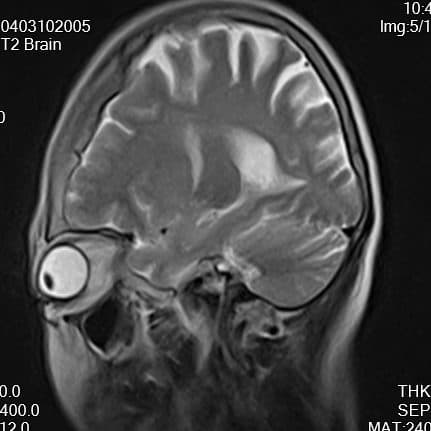
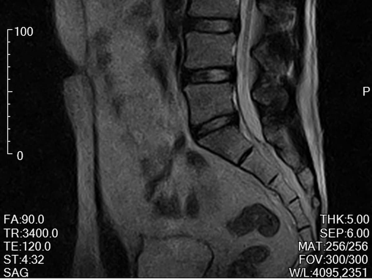
Clinical images require written authorization from the source institution and removal of patient identifiers.
Talk to us about your site.
Tell us where the system needs to go and what you need to image. We will tell you what is possible.