Skip to content
PANTORISMRI

Products

Two branches. One architecture.

Human clinical imaging and veterinary imaging share the same magnet, noise-cancellation and reconstruction platform. What changes is the coil set, the protocol and the workflow.

Branch one

Human clinical imaging.

Extremity, head and whole-body systems, each installable in an ordinary room.

0.25T · Cleared in China and the United States

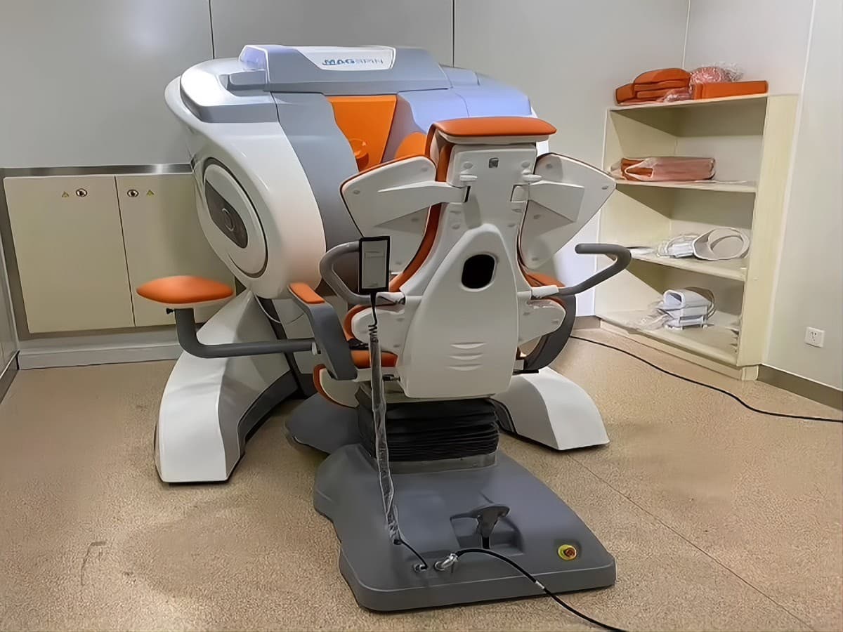
MSK & Extremity MRI

PANTORIS Formula III

Weight-bearing imaging for orthopedics, sports medicine and joint care.

  • Standing / seated scanning close to the real load-bearing state
  • Reduced metal-artifact interference — usable for post-operative follow-up
  • Small modular enclosure — about 12 m² — plus single-phase power
  • 0.25T C-type open permanent magnet — 24 mT/m, 80 mT/m/ms, under 5 kW
  • Three dedicated coils: knee/ankle, wrist/elbow and shoulder
View detailsIn-clinic · Mobile & trailer · Upper floors
Sample unit completed

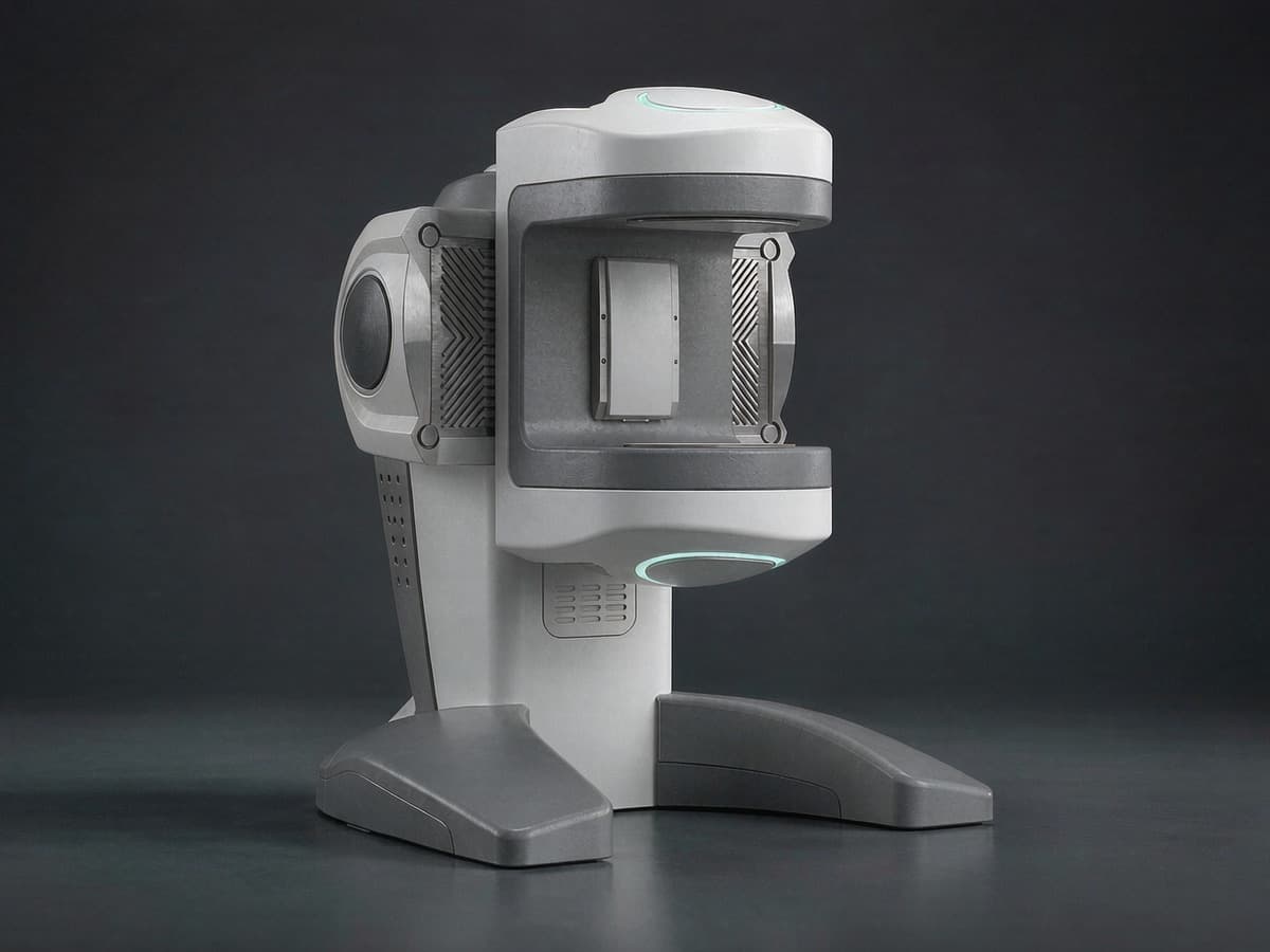
Head & Neuro MRI

PANTORIS Formula II

Head imaging at the bedside and in the operating room.

  • Designed for ward and OR environments
  • Brings imaging to the patient instead of moving the patient to imaging
  • Research collaboration with a Hong Kong-based research institute
View detailsIn-clinic · Bedside · Intraoperative

0.2T · Sample unit completed, registration in progress

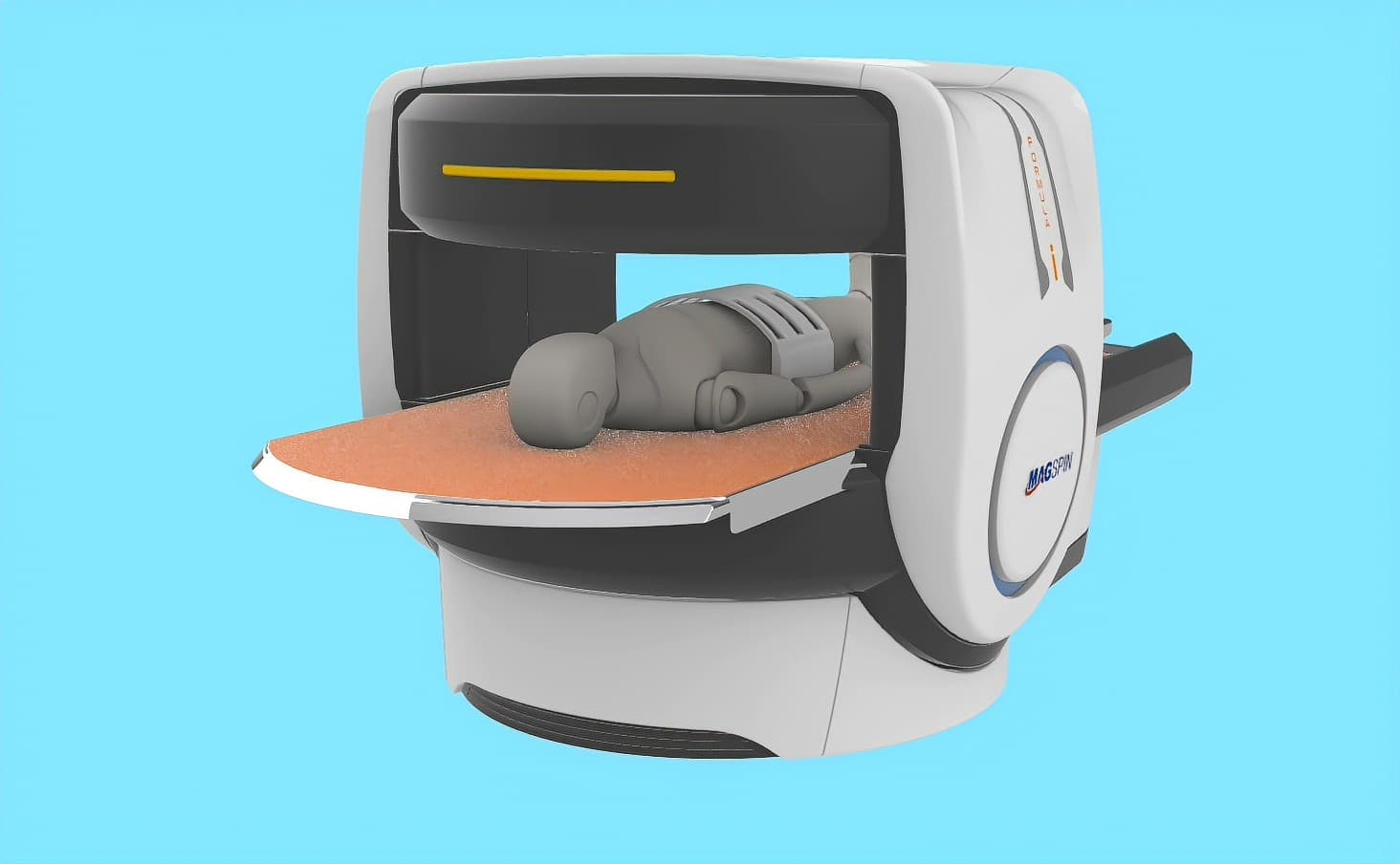
Whole-Body MRI

PANTORIS Formula I

Multi-region scanning in a system that travels.

  • Multi-region coverage in one system
  • AI-assisted reporting workflow
  • Trailer-mountable and installable on upper floors
View detailsIn-clinic · Mobile & trailer · Upper floors
Branch two

Veterinary imaging.

Equine and small-animal systems. In veterinary use, no medical device registration applies.

0.3T · No medical device registration required for veterinary use

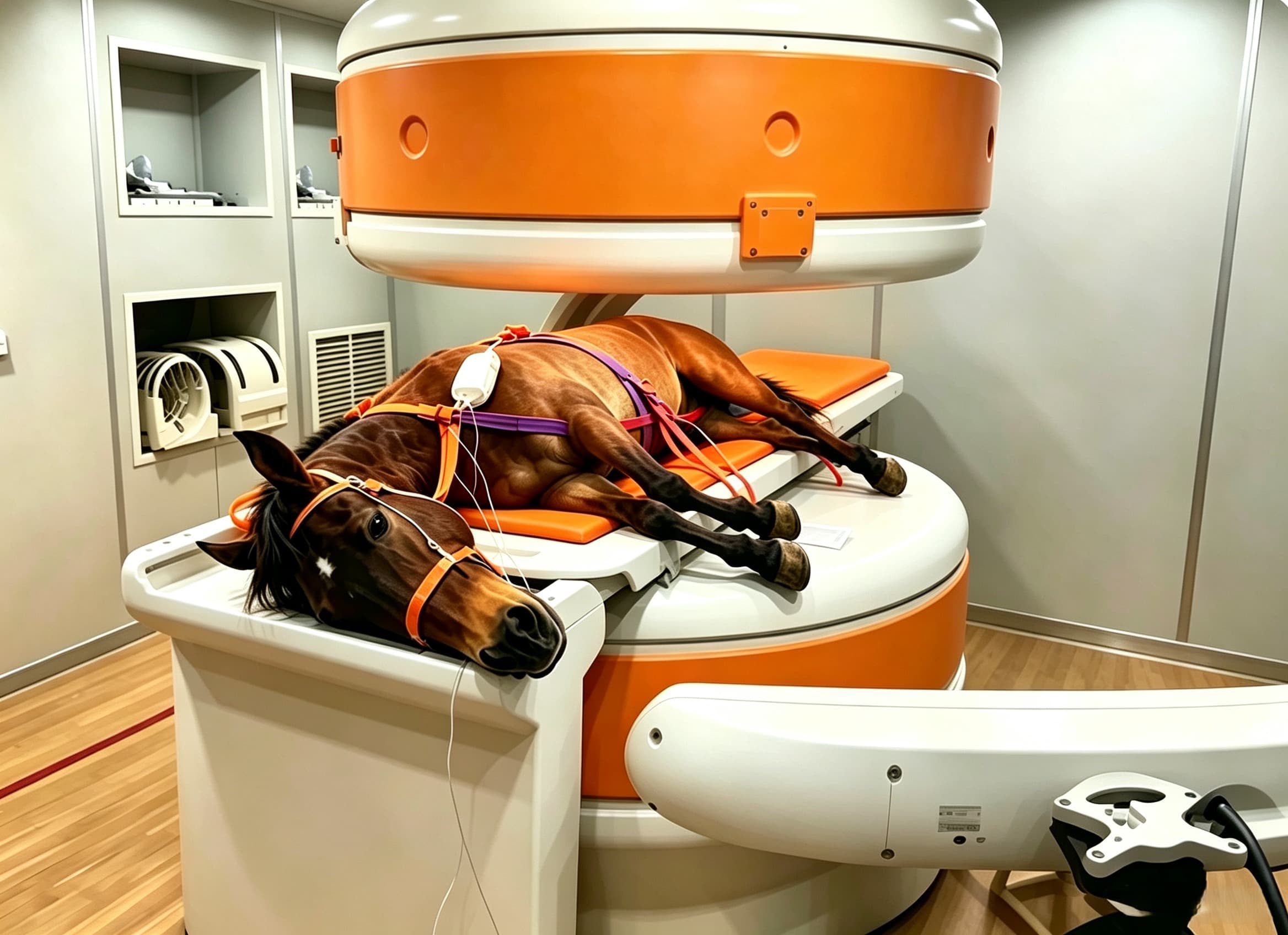
Veterinary MRI

PANTORIS Veterinary Series

Equine and small-animal imaging, from the hoof to the head.

  • Standing sedation scanning for horses — avoids the risk of general anaesthesia
  • Dedicated hoof and fetlock coils for the distal limb, where X-ray and ultrasound fall short
  • Two configurations: 0.3T U-shape with a 330 mm gap, or 0.25T double-column with 300 mm
  • Covers animals up to 200 kg, FOV 100–300 mm, DICOM 3.0 with PACS and patient worklist
View detailsEquine clinic · Mobile & trailer

Not sure which configuration fits?

Tell us the anatomy you need to image and the room you have. We will tell you which system fits and what the siting requires.